











In modern clinical oncology, measuring tumor progression from medical imaging is a routine yet labor-intensive task. Typically, radiologists mark a tumor on a computed tomography (CT) scan by drawing a straight line across its longest visible diameter, a standard known as a RECIST (Response Evaluation Criteria in Solid Tumors) measurement. While efficient for tracking treatment response in two dimensions, this single line falls short of capturing a tumor’s complete three-dimensional shape. Creating a full 3D segmentation requires painstakingly outlining the mass across every individual slice where it appears, demanding significant time and clinical expertise.
To bridge the gap between rapid 2D measurements and comprehensive 3D analysis, researchers developed Lumina, an automated AI system designed to generate a complete 3D tumor segmentation mask using nothing more than a CT scan and a single RECIST line. Engineered specifically for the competitive landscape of the FLARE 2026 pan-cancer segmentation challenge, the system operates under strict computational parameters: it is built to execute entirely on a CPU within an 8 GB memory limit, maintaining an inference window of under 60 seconds per case.
Understanding What Lumina Does
The core premise behind Lumina is to simplify the complex challenge of volumetric tumor segmentation by leveraging existing clinical workflows. When a radiologist performs a RECIST measurement, they establish a 2D diameter marker across the longest axis of the tumor. Although this indicator communicates the location and approximate size of the target lesion, it lacks volumetric depth.

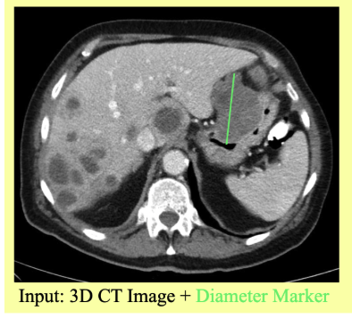
Lumina uses this sparse prompt to guide a specialized 3D segmentation model. Instead of forcing an algorithm to autonomously scan an entire abdomen or thorax to identify every potential abnormality, the system receives a precise geographic hint. The input pipeline processes a 3D CT scan alongside a 2D RECIST line marking one specific tumor. In return, the model outputs a precise 3D mask of that exact tumor, assigning an individual label to every relevant voxel—the three-dimensional equivalent of a pixel. By narrowing the scope of the problem around the RECIST prompt, the system reduces ambiguity and focuses its computational power entirely on defining the boundaries of the targeted mass.
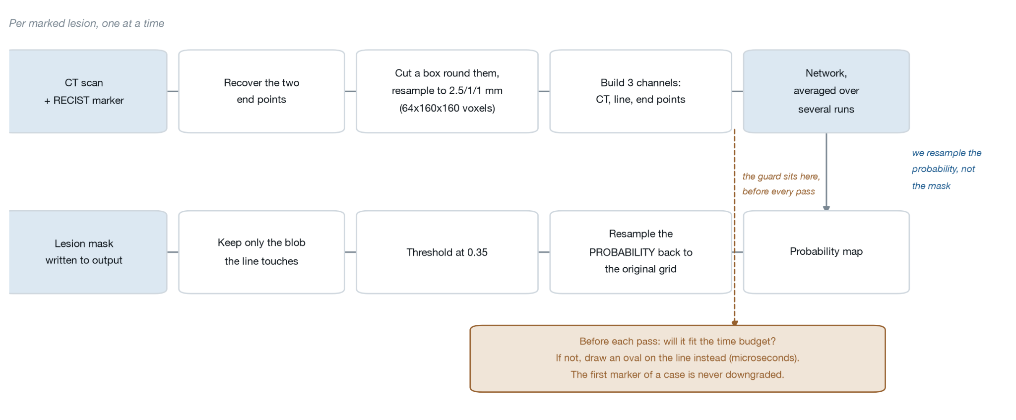
The architecture of the Lumina pipeline relies on a carefully orchestrated sequence of data transformations. The process begins with the raw 3D CT scan and the accompanying RECIST marker line. The system converts this line into additional input channels that encode spatial location and endpoint parameters, crops and resamples the image data to a uniform grid, and passes the resulting three-channel input through a deep 3D segmentation network. The network subsequently generates a probability map, which undergoes resampling, thresholding, and connected-component selection to yield the final 3D tumor mask.
Navigating Data Preprocessing and Metadata Realities
Developing the system required careful inspection of the underlying data structures, which were provided in compressed NumPy array formats. Each file contained volumetric CT scans, marker lines, voxel spacings, spatial origins, coordinate directions, and, in training files, ground-truth segmentations. Before writing model code, the development team analyzed image values and spatial metadata, uncovering critical operational nuances.
A primary consideration involved the intensity values of the scans. While CT images traditionally store data in Hounsfield units—where water registers near zero and dense bone exceeds one thousand—the dataset had already been preprocessed into a fixed zero-to-255 brightness range. Because the original Hounsfield units were unavailable, the team bypassed standard CT windowing techniques and instead analyzed the actual intensity distribution of the provided files. Notably, more than half of all voxels possessed a value of exactly zero, representing air surrounding or within the body.
Coordinate ordering also proved vital. Discrepancies between spatial metadata storage formats and internal array indexing can easily introduce geometric errors. The spacing array stored dimensions in conventional spatial order, whereas NumPy arrays rely on a different indexing sequence. To prevent spatial inaccuracies, the pipeline converted these coordinates uniformly at the input boundary and maintained a strict internal convention throughout all subsequent processing steps. This underscored a broader lesson for medical imaging development: avoiding assumptions that data will automatically adhere to conventions established in other datasets or external tutorials.
Translating RECIST Lines into Neural Network Inputs
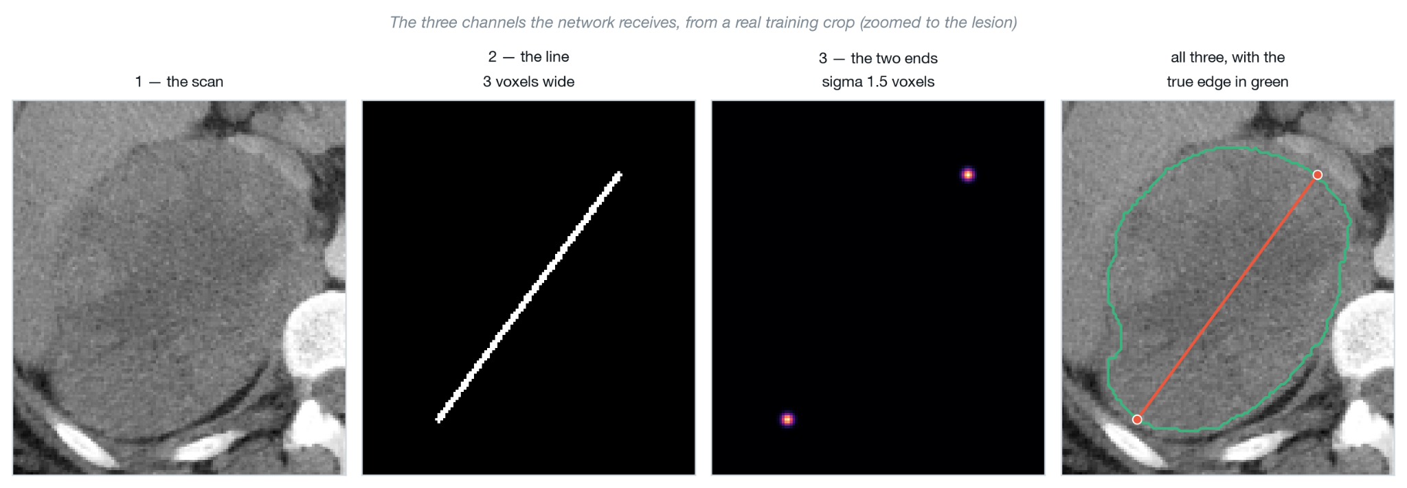
To make use of a RECIST line, a neural network requires a structured multi-channel input format. Lumina dedicates its first input channel to the CT scan itself, while the remaining channels represent the RECIST line and its geometric properties in a format accessible to the network.
Rather than relying solely on a thin binary trace of the line, the system incorporates the exact endpoints of the diameter measurement. These endpoints define the measured extent of the tumor and supply the network with critical context regarding its physical scale and orientation. This endpoint information is translated into the network using Gaussian functions, where values are elevated near the endpoint and gradually taper off over distance.
Calculating these Gaussian distributions locally around each endpoint prevents unnecessary computational overhead. Because the influence of the Gaussian function drops sharply within a short distance, computing it over the entire volumetric space would waste resources. Furthermore, the RECIST line itself is redrawn directly from its endpoints at the exact resolution required by the network. Resampling a pre-existing thin line from a different resolution risks thinning or fracturing the line, whereas regenerating it ensures consistency with the underlying image grid.
Aligning Tumors on a Common Spatial Grid
Because CT scans can originate from different scanners with varying slice thicknesses and resolutions, voxel spacings fluctuate widely across medical datasets. A specific number of voxels in one scan may represent a completely different physical volume than the same count in another. To establish uniformity, the system extracts a fixed-size crop for each marked tumor and resamples it to a standardized voxel spacing.
The pipeline utilizes a target spacing closely aligned with the median slice spacing found in the training dataset, alongside a standardized spatial crop derived from the distribution of lesion sizes. This configuration ensures the network consistently receives inputs of identical dimensions. Intensity normalization is similarly standardized using mean and standard deviation statistics computed strictly from the training data. By locking in these normalization parameters and applying them uniformly across validation and test sets, the pipeline prevents data leakage and preserves the integrity of the evaluation process.
Constructing the 3D Segmentation Network and Loss Function
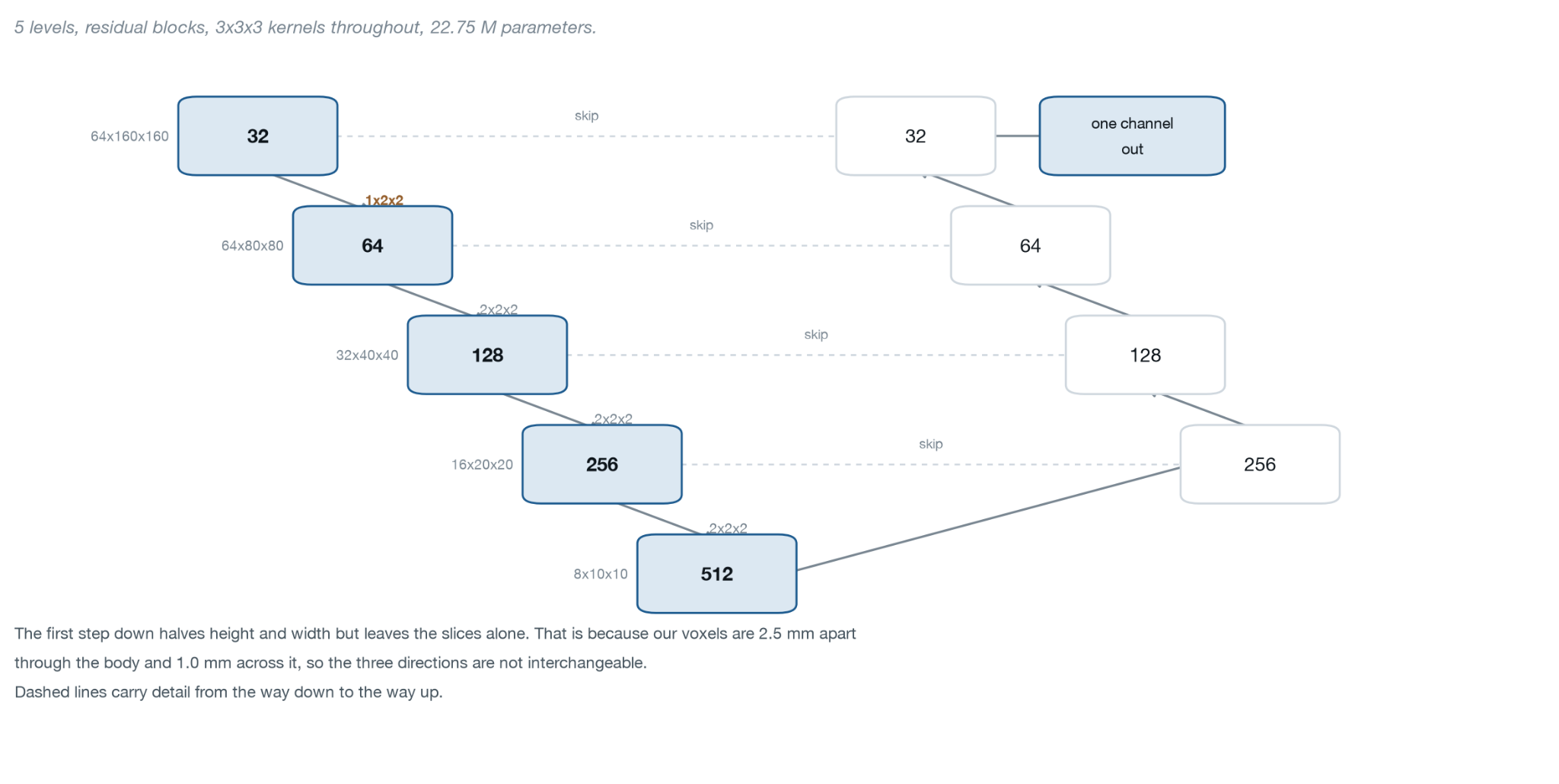
At the heart of Lumina lies DynUNet, a robust 3D convolutional neural network architecture built upon foundational U-Net principles. The network features an encoder-decoder structure augmented by skip connections that bridge shallow and deep layers. While the encoder captures broad contextual information by reducing spatial dimensions, the decoder reconstructs the fine details necessary for accurate boundary delineation. Skip connections pass high-resolution spatial details directly from the encoder to the decoder, preventing the loss of critical structural boundaries during downsampling.
The network processes three input channels simultaneously: the CT scan, the mapped RECIST line, and the endpoint heatmap blobs. Due to the anisotropic nature of medical scans—where inter-slice spacing often exceeds in-plane resolution—the network’s downsampling strides are carefully controlled. Specifically, the network preserves the depth dimension during the initial downsampling step to retain inter-slice detail rather than discarding it prematurely.
Training an effective segmentation model also requires a specialized loss function capable of guiding the network toward precise boundary alignment. Lumina combines Dice loss, which emphasizes overall volumetric overlap between the predicted and reference regions, with binary cross-entropy, which operates at the individual voxel level to penalize incorrect probability estimates. To address the strict boundary tolerances required by clinical evaluations, the team introduced a specialized boundary-band loss term. By isolating a thin shell around the ground-truth surface via morphological operations, the network receives targeted feedback specifically penalizing surface errors, significantly improving segmentation performance on larger, complex lesions.
Post-Processing and Probability Mapping
Once the segmentation network generates its initial probability map—where every voxel holds a value between zero and one representing its likelihood of belonging to the tumor—the pipeline initiates a structured post-processing sequence.
Instead of thresholding the probability map immediately at the network’s internal resolution, the system first resamples the probability map back onto the original coordinate grid of the input CT scan. Resampling the probability values prior to thresholding allows for smoother interpolation across the original image space, resulting in cleaner and more accurate final contours. Following this resampling step, the system applies a validated threshold of 0.35 to generate a binary segmentation mask.

To eliminate extraneous tissue regions or disconnected artifacts that frequently appear during thresholding, the pipeline uses the original RECIST line to identify and isolate the correct connected component. The system retains the specific volumetric component that intersects with the RECIST marker line as the final tumor mask. If an edge case arises where the line does not directly intersect a component, a fallback mechanism selects the component whose centroid lies closest to the midpoint of the line.
Operating Within Strict CPU and Memory Constraints
Operating within the strict performance constraints of an 8 GB memory limit and a 60-second inference ceiling required deliberate hardware and algorithmic optimizations. Because multi-threaded CPU operations can introduce microscopic numerical variances that alter threshold outcomes near boundary cutoffs, the pipeline explicitly pins the PyTorch thread count to a fixed value. This guarantees numerical reproducibility across different execution runs.
To maximize segmentation accuracy without violating runtime limits, the system employs an adaptive ensemble strategy. Ensembling combines predictions from multiple model passes—including flipped image orientations and alternative architectures—to yield more robust segmentations. However, because scans can feature variable numbers of marked tumors, running an exhaustive ensemble on every lesion in a complex scan would easily exceed the 60-second limit. Lumina dynamically budgets its inference passes based on the lesion count, scaling down the number of ensemble passes as the quantity of marked tumors increases. This maintains predictable execution times while preserving high segmentation quality.
Evaluation Results and Clinical Insights
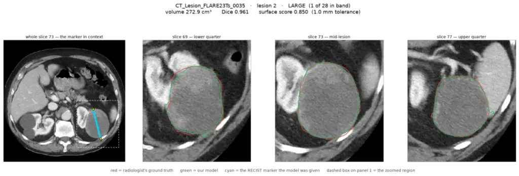
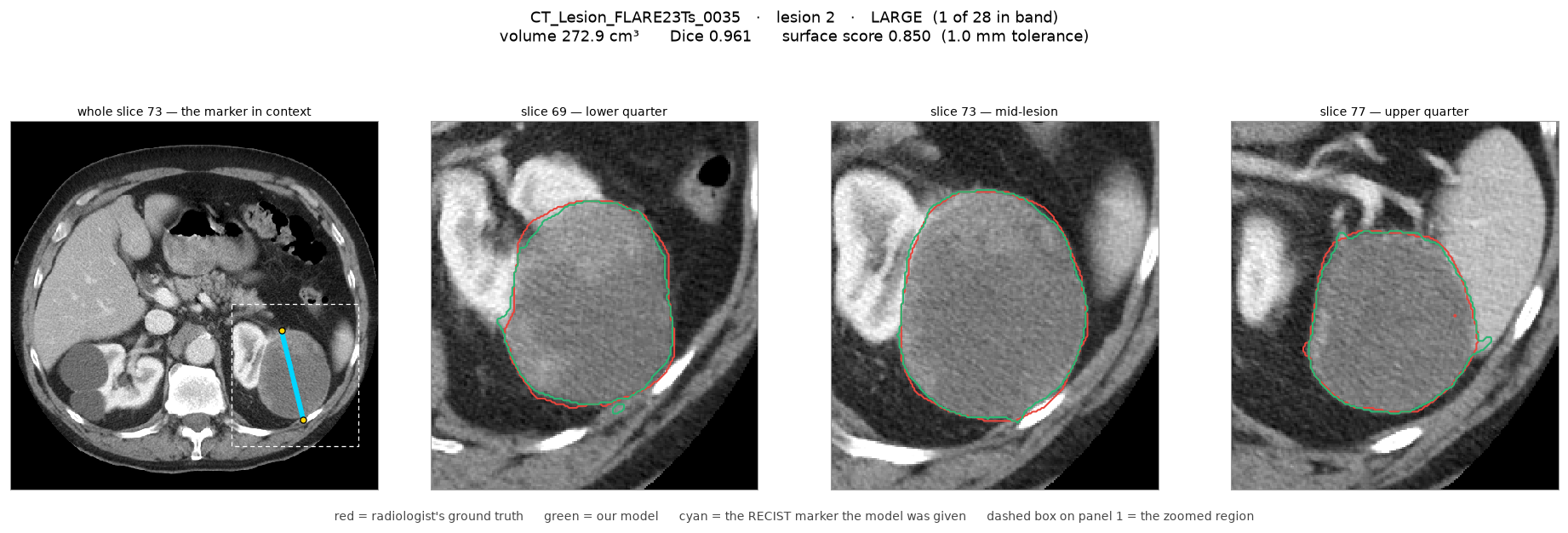
When evaluated on 217 held-out test scans, Lumina demonstrated robust performance, achieving a strong Dice similarity coefficient and surface distance metrics under strict clinical tolerances. The median inference time per case remained efficient, while peak memory consumption stayed comfortably within the allocated container limits. Qualitative reviews conducted alongside radiologists offered further context into the system’s behavior across different lesion types.
Analysis revealed that while well-circumscribed lesions with sharp boundaries yielded high geometric agreement, lesions characterized by indistinct margins or tissue textures blending seamlessly into surrounding anatomy presented greater challenges. Radiologist reviews emphasized that numerical metrics alone do not always capture clinical validity, as even expert human annotators can experience ambiguity when defining complex tumor borders.
Ultimately, the development of Lumina illustrates a practical, resource-conscious pathway for transforming rapid 2D clinical markings into comprehensive 3D volumetric data. By combining targeted input prompts, fixed spatial normalization, boundary-aware loss functions, and disciplined CPU inference strategies, the system offers a scalable blueprint for automated medical image segmentation under strict computational boundaries.